

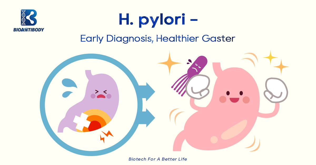
Helicobacter pylori (HP) is a bacterial that lives in the stomach and adheres to the gastric mucosa and intercellular spaces, causing inflammation. HP infection is one of the most common bacterial infections, infecting billions of people worldwide. They are the leading cause of ulcers and gastritis (inflammation of the stomach lining).
High infection in children and familial aggregation are the significant characteristics of HP infection, and family transmission may be the main route HP infection is a major causative factor in chronic active gastritis, peptic ulcer, gastric mucosa-associated lymphoid tissue (MALT) lymphoma, and gastric cancer. In 1994, the World Health Organization/International Agency for Research on Cancer (WHO/IARC) designated Helicobacter pylori as a class I carcinogen
Gastric mucosa – the body armor of the stomach
Under normal circumstances, the stomach wall has a series of perfect self-protection mechanisms (the secretion of gastric acid and protease, the protection of insoluble and soluble mucus layers, regular exercise, etc.), which can resist the invasion of thousands of microorganisms that enter by mouth.
HP has independent flagella and a unique helical structure, which not only plays an anchoring role during bacterial colonization, but also can become spherical and form a self-protecting morphology in harsh environments. At the same time, Helicobacter pylori can produce a variety of toxins, which determine that Helicobacter pylori can pass through the gastric juice layer through its own power and resist gastric acid and other unfavorable factors, becoming the only microorganism that can survive in the human stomach.
Pathogenesis of Helicobacter pylori
1. Dynamic
Studies have shown that Helicobacter pylori has a strong ability to move in a viscous environment, and the flagella is necessary for the bacteria to swim to the protective mucus layer on the surface of the gastric mucosa.
2. Endotoxin-associated protein A (CagA) and vacuolar toxin (VacA)
Cytotoxin-associated gene A (CagA) protein secreted by HP can trigger local inflammatory response. CagA-positive Helicobacter pylori infection can also significantly increase the risk of atrophic gastritis, intestinal metaplasia and gastric cancer.
Vacuolating cytotoxin A (VacA) is another most important pathogenic factor of Helicobacter pylori, which can enter mitochondria to regulate the function of organelles.
3. Flagellin
Two flagellin proteins, FlaA and FlaB, constitute the major components of flagellar filaments. Changes in flagellin glycosylation affect strain motility. When the level of FlaA protein glycosylation was increased, both the migratory ability and the colonization load of the strain increased.
4. Urease
Urease generates NH3 and CO2 by hydrolyzing urea, which neutralizes gastric acid and raises the pH of surrounding cells. In addition, urease participates in inflammatory responses and promotes adhesion by interacting with CD74 receptors on gastric epithelial cells.
5. Heat shock protein HSP60/GroEL
Helicobacter pylori absorbs a series of highly conserved heat shock proteins, of which co-expression of Hsp60 with urease in E. coli greatly increases urease activity, allowing the pathogen to adapt and survive in the hostile ecological niche of the human stomach.
6. Hook-related protein 2 homolog FliD
FliD is a structural protein that protects the tip of flagella and can repeatedly insert flagellin to grow flagellar filaments. FliD is also used as an adhesion molecule, recognizing glycosaminoglycan molecules of host cells. In infected hosts, anti-flid antibodies are markers of infection and can be used for serological diagnosis.
Test Methods:
1. Stool test: The stool antigen test is a non-invasive test for H. pylori. The operation is safe, simple and fast, and does not require oral administration of any reagents.
2. Serum antibody detection: When Helicobacter pylori infection occurs in the body, the human body will have anti-Helicobacter pylori antibodies in the blood due to an immune response. By drawing blood to check the concentration of Helicobacter pylori antibodies, it can reflect whether there is Helicobacter pylori in the body. bacterial infection.
3. Breath test: This is a more popular inspection method at present. Oral urea containing 13C or 14C, and breath test the concentration of carbon dioxide containing 13C or 14C after a period of time, because if there is Helicobacter pylori, urea will be detected by its specific urea. The enzymes break down into ammonia and carbon dioxide, which is exhaled from the lungs through the blood.
4. Endoscopy: allows careful close observation of gastric mucosal features such as redness, swelling, nodular changes, etc.; endoscopy is not suitable for patients with severe complications or contraindications and additional costs (anesthesia, forceps) ).
Bioantibody related products of H. pylori recommendations:
H. Pylori Antigen Rapid Test Kit (Lateral Chromatography)
H. Pylori Antibody Rapid Test Kit (Lateral Chromatography)
Post time: Oct-18-2022